
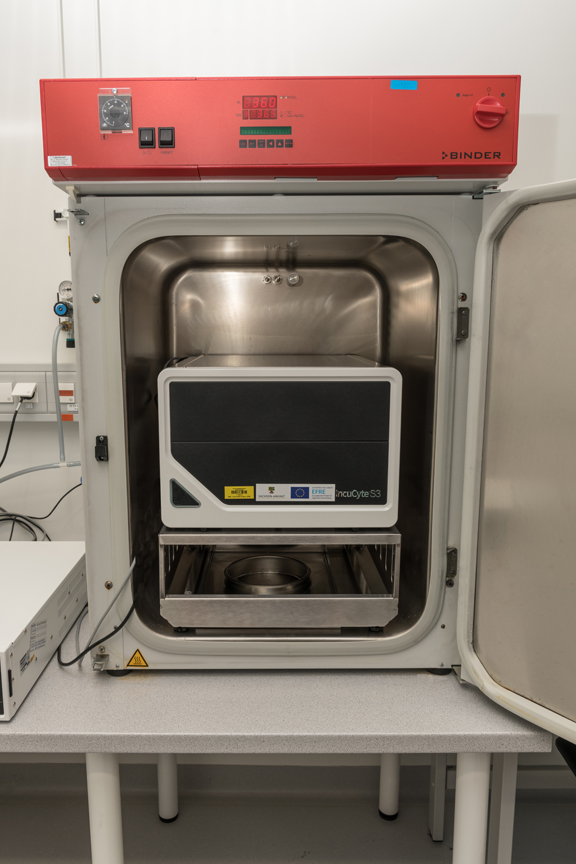
- High-throughput live cell analysis system for long-term experiments
- Bright field, phase contrast and fluorescence images
- Objectives 4x, 10x and 20x
- Existing analysis options for: growth and cell toxicity assays, migration/scratch, spheroids, T-cell assays, angiogenesis, chemotaxis and neuronal differentiation
- It is possible to carry out necessary cell culture steps on site
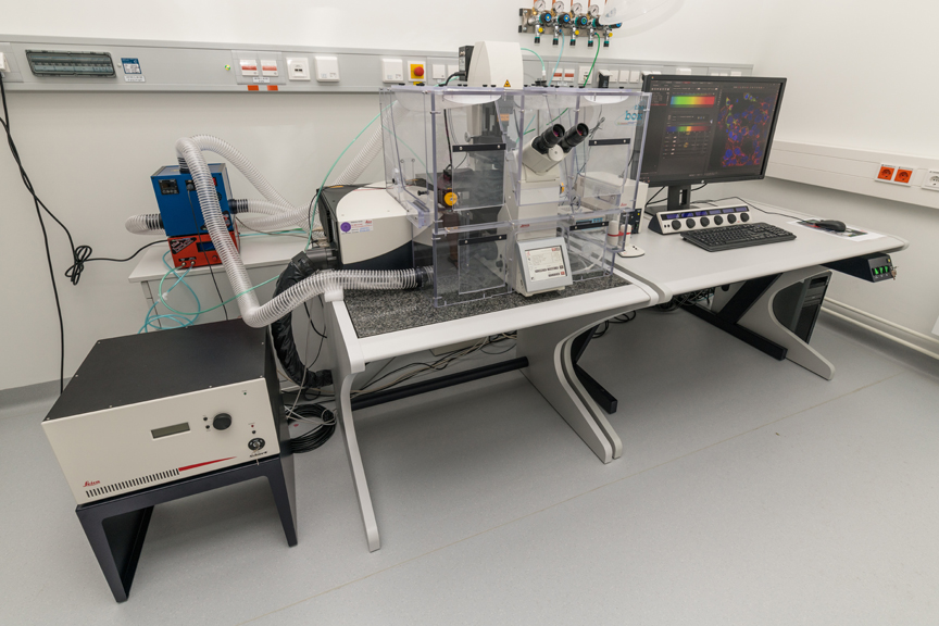
- Laser: pulsed white light laser (470 nm - 670 nm), diode (405 nm), argon (458 nm, 476 nm, 488 nm & 496 nm)
- Tandem scanner: conventional scanner (10-1400 Hz) and resonance scanner (8MHz)
- Detectors: 2x HyD, 2x PMT incl. time gating and photon counting
- Objectives: 20x - 60x with different immersion (dry, oil, glycerin and water incl. water pump)
- Ludin cube incubation system for live cell analysis
- Automated high content screening software (Leica HCS A)
- HyVolution2
- Cuda workstation and off-station including navigator, 3D visualization and Hyugens deconvolution
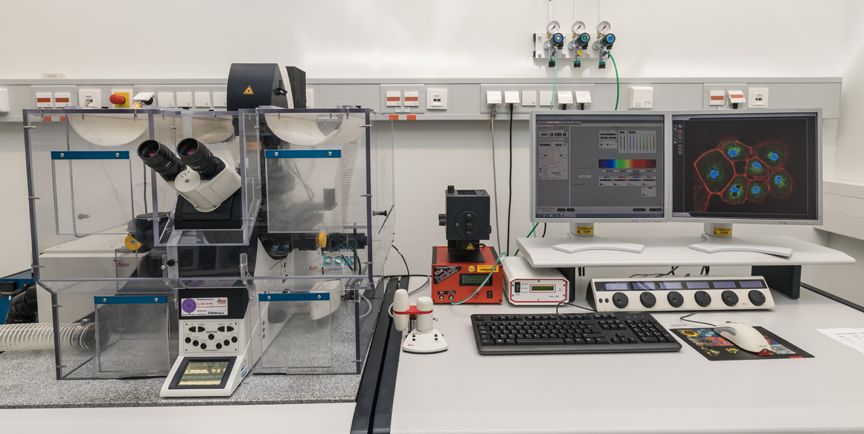
- Laser: Pulsed white light laser (470 nm - 670 nm) as well as diode (405 nm), argon (458 nm, 476 nm, 488 nm & 496 nm), DPSS561 (561 nm) and HeNe633 (633 nm)
- Tandem scanner: conventional scanner (10-1400 Hz) and resonance scanner (8MHz)
- Detectors: 2x HyD and 3x PMT incl. time gating and photon counting
- automated high content screening software (Leica HCS A) with DFC 360 FX camera (40 MHz)
- Ludin cube incubation system for live cell analysis
- FRET and FRAP software applications
- Objectives: 20x - 100x with different immersion media (oil, dry, water)
- 3D deconvolution and colocalization application
- X/Y/Z motorized inverted epi-fluorescence microscope with 3D imaging option
- Multi-positioning and stitching
- Perfect Focus System (PFS)
- Pecon-liveCell incubation chamber with temperature and CO2 control
- 12 MP color camera (cooled)
- X-Cite illumination with adjustable intensity from UV to infrared
- Available objectives: 4x, 10x, 20x and 63x Plan-Apo, 100x TIRF
- Available filters (motorized): DAPI, CFP, GFP, YFP, RFP, Cy5, Cy7
- Deconvolution and real-time deconvolution equipment for noise reduction and high resolution
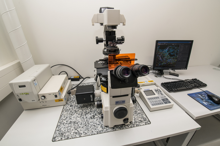
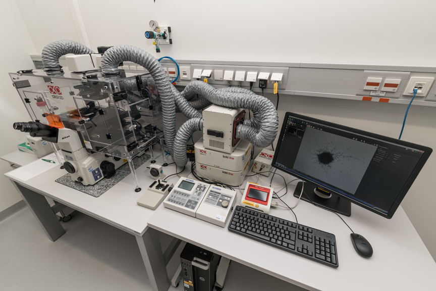
- Z-Motorized Inverted Epi-Fluorescence microscope with 3D imaging option
- DS-1QM camera with high sensitivity, which enables recordings even in the infrared range with high resolution and high recording speeds (15 frames / sec).
- 5 MP color camera (cooled)
- X-Cite illumination with adjustable intensity from UV to infrared
- Available objectives: 4x, 10x, 20x and 63x plan apo, 100x TIRF
- Available filters (motorized): DAPI, CFP, GFP, YFP, RFP, Cy5, Cy7
- Deconvolution and real-time deconvolution equipment for noise reduction and high resolution
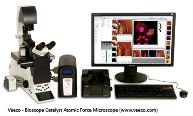
- Bioscope Catalyst combines atomic force and fluorescence microscopy (Leica System)
- Atomic force measurements can be combined with optical data
- System and application details can be found here
- The Core Facility Imaging only offers experienced users access to this special system
This device is located at the Medical Campus Steintor (Grosse Steinstr. 52) - Department for Anatomy and Cell Biology.
Contact:
Dr. rer. nat. Tim Hohmann (Tel.:0345 557 1316)
Prof. Dr. med. Faramarz Dehghani (Tel.:0345 557 1707)
- inverse confocal system with solid state lasers
- different objectives with 10x, 20x, 40x and 63x magnification
- PMT detector for sequential image acquisition
This device is located at the Medical Campus Steintor (Grosse Steinstr. 52) - Department for Anatomy and Cell Biology.
Contact:
Dr. rer. nat. Tim Hohmann (Tel.:0345 557 1316)
Prof. Dr. med. Faramarz Dehghani (Tel.:0345 557 1707)